When JDM patients complain of pain in the lower extremities, pediatric rheumatologists may initially consider this to be an exacerbation of myositis.
#X RAY STRESS FRACTURE SHIN SKIN#
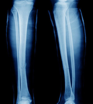
Juvenile dermatomyositis (JDM) is a rare chronic inflammatory disease of childhood, characterized by skin involvement and proximal muscle weakness. In addition to preventing JDM patients from getting osteoporosis, we need to consider stress reactions when children with JDM complain of sudden shin pain. Their underlying disease and weakness, treatment with glucocorticoid and methotrexate, or inactivity may have resulted in these tibial injuries, and made these patients more predisposed than other children. We experienced 6 children with JDM who presented with shin pain, and who were diagnosed with tibial stress fractures or stress reactions. In all 6 patients, the tibial stress injuries improved with only rest and/or analgesics. In one patient, we could not find any abnormalities in his radiograph, but the subsequent MRI showed tibial stress reaction. They were diagnosed with tibial stress injuries based on a combination of radiographs, three-phase bone scans, and magnetic resonance imaging (MRI), and 5 out of 6 patients had been treated with prednisone and/or methotrexate at onset of tibial stress injuries.

Case presentationĪll 6 patients with JDM presented with shin pain or tenderness in the anterior tibia without any evidence of excessive exercise or traumatic episode. Here we report 6 JDM patients who presented with shin pain, and the imaging appearance of tibial stress fractures or stress reactions. They sometimes occur in patients with pathological conditions of bone metabolism such as osteoporosis or rheumatoid arthritis, but there are previously no cases reported in juvenile dermatomyositis (JDM). (2-4) The Talar Bump Test may help differentiate tibial stress fracture from MTSS.Tibial stress injuries are frequent injuries of the lower extremity and the most common causes of exercise-induced leg pain among athletes and military recruits. (1) Single leg hopping is painful in about half of MTSS cases (and 70-100% of stress fractures). Applying a vibrating tuning fork over the tibia may help detect stress fracture with 75% sensitivity. More focal tenderness, the presence of anterior tibial tenderness, or any significant swelling suggests stress fracture. Tenderness from MTSS should involve at least 5 cm of the tibial border. Prolonged stress may generate a periosteal reaction detectable as a “rough” or “bumpy” feel upon palpation. Pain that persists more than five minutes post-activity carries a higher suspicion of stress fracture.Ĭlinical evaluation demonstrates diffuse tenderness over the posteromedial tibial border.

Initially, symptoms may subside during training, but as the condition progresses (toward stress fracture), symptoms may linger throughout activity or even at rest. Symptoms are often worse with exertion – particularly at the beginning of a workout. The clinical presentation of MTSS includes vague, diffuse pain over the middle to distal posteromedial tibia. Prolonged insult may lead to a tibial stress fracture, and many authors now believe that MTSS and stress fracture represent two different points along a continuum of bony stress reaction. Stress reactions occur when the normal adaptive remodeling response cannot keep pace with excessive training loads, i.e., high demands with inadequate recovery times. Healthy bone responds to this stress by remodeling itself more densely. The stress of exercise can temporarily weaken bone. Research suggests that traction periostitis may be an inflammatory precursor to a tibial stress fracture.


Early etiological theories focused on myofascial strain, but current evidence shows that a bony stress reaction is the most likely cause of MTSS.


 0 kommentar(er)
